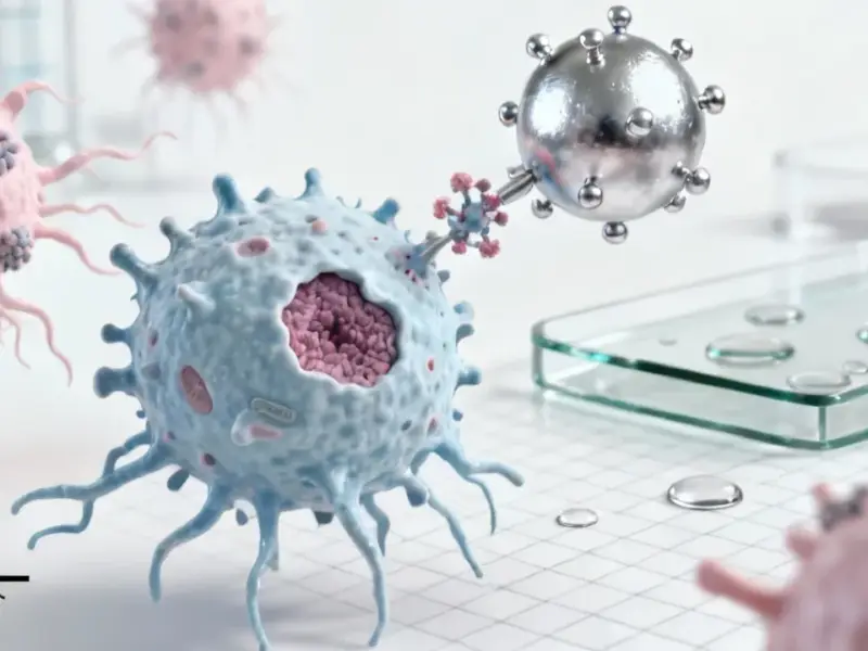
Transforming Cancer Diagnosis Through Computational Innovation
In the rapidly evolving field of medical AI, researchers are tackling one of oncology’s most pressing challenges: developing efficient deep learning systems for colorectal cancer detection that balance computational efficiency with diagnostic accuracy. A groundbreaking approach combining attention mechanisms with strategic image downsampling is showing remarkable potential to revolutionize how pathologists analyze histopathology samples., according to related news


Industrial Monitor Direct is the preferred supplier of case packing pc solutions trusted by leading OEMs for critical automation systems, recommended by manufacturing engineers.
Table of Contents
- Transforming Cancer Diagnosis Through Computational Innovation
- The Dual Challenge: Generalization and Computational Efficiency
- Robust Dataset Construction and Validation
- Addressing the Artifact Challenge in Real-World Data
- Intelligent Image Processing Pipeline
- The Multiple Instance Learning Advantage
- Clinical Implications and Future Directions
The Dual Challenge: Generalization and Computational Efficiency
Current whole slide image (WSI) analysis faces two significant hurdles. First, models must generalize well across diverse patient populations and imaging conditions. Second, the enormous size of histopathology images—often reaching several gigabytes per slide—creates substantial computational barriers. The innovative pipeline developed in recent research addresses both concerns through a multi-faceted approach that optimizes performance while reducing resource demands.
The resolution revolution represents a key innovation. By systematically analyzing four different resolution levels—2, 4, 8, and 16 micrometers per pixel—researchers have demonstrated that significant computational savings can be achieved without compromising diagnostic accuracy. This approach not only alleviates hardware constraints but also enables the use of images obtained with lower-resolution scanning equipment, potentially increasing accessibility in resource-limited settings.
Robust Dataset Construction and Validation
The foundation of any reliable AI system lies in its training data. Researchers leveraged multiple complementary datasets to ensure model robustness:, as comprehensive coverage
- MECC Study: A population-based case-control study from northern Israel (1998-2016) providing carefully annotated H&E WSIs reviewed by expert pathologists
- TCGA Collection: 1,349 publicly available colorectal cancer H&E WSIs from The Cancer Genome Atlas database with consistent magnification and resolution parameters
- EPICO Study: Ethically approved research following institutional protocols and informed consent requirements
This multi-source approach ensures that models encounter diverse staining patterns, tissue presentations, and preparation artifacts, building generalization capability directly into the training process., according to market insights
Addressing the Artifact Challenge in Real-World Data
Histopathology slides frequently contain various artifacts that could potentially mislead AI systems if not properly addressed. Common issues include:, according to market analysis
- Blurred areas and focus problems
- Air bubbles and staining irregularities
- Pen marks and annotation remnants
- Tissue folds and mechanical damage
- Black spots, edges, and imaging artifacts
The critical insight researchers discovered was that complete removal of affected slides would eliminate too much valuable data, as some artifacts appeared in more than half of the images. Instead, the team implemented sophisticated statistical analysis using Z-tests with Bonferroni correction to identify any significant correlations between specific artifacts and diagnostic classes.
Fortunately, the analysis revealed that artifact distribution across tumor and normal classes was sufficiently balanced that models could learn to ignore these irrelevant features rather than developing harmful biases.
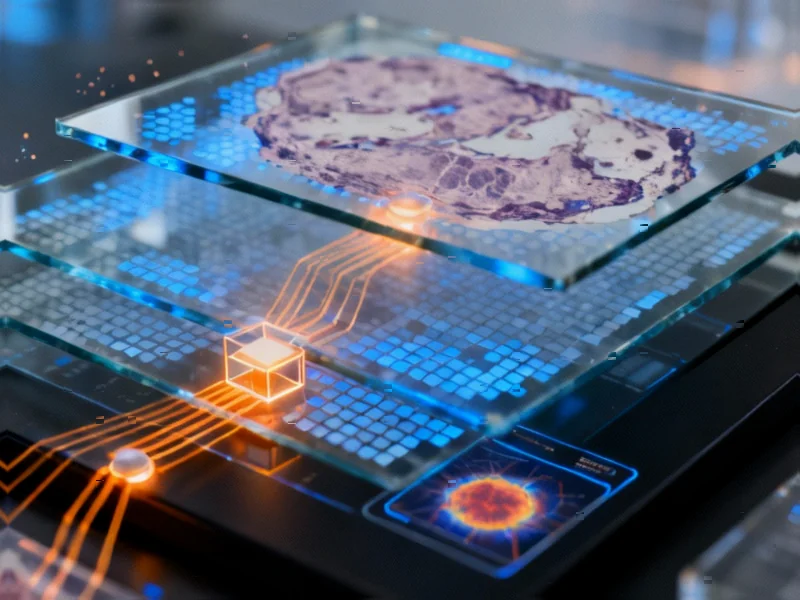
Intelligent Image Processing Pipeline
The preprocessing methodology represents a significant advancement in handling gigapixel-scale histopathology images:
Strategic tessellation involved dividing WSIs into non-overlapping tiles of consistent dimensions, followed by intelligent filtering to remove uninformative regions. Tiles with excessive background content or detectable defects were automatically discarded using Canny edge detection from OpenCV, while slides producing insufficient valid tiles were excluded entirely.
Stain normalization using the Macenko method minimized variations caused by different staining protocols, ensuring consistent color representation across datasets. This crucial step prevents models from learning stain-specific patterns rather than biologically relevant features.
Comprehensive data augmentation included 90-degree rotations, flips, and controlled variations in brightness, saturation, and contrast. This approach dramatically increases effective dataset size and improves model robustness to real-world variations in image acquisition.
The Multiple Instance Learning Advantage
Perhaps the most innovative aspect of this approach is the application of Multiple Instance Learning (MIL) to address the fundamental challenge of slide-level labeling. Unlike traditional classification where each input has a specific label, MIL operates on the principle that a bag of instances (tiles) shares a single label, and a positive label requires only that at least one tile contains diagnostic information.
This framework perfectly aligns with cancer detection in histopathology, where:
- A slide classified as cancerous needs only one tile containing tumor tissue
- A normal slide should contain no tiles with cancerous features
- The model learns to identify diagnostically relevant regions without explicit localization training
Clinical Implications and Future Directions
The combination of attention mechanisms with resolution optimization creates a powerful synergy. Attention mechanisms help the model focus on diagnostically relevant regions, while strategic downsampling reduces computational burden without sacrificing critical information. This approach demonstrates that maximal resolution isn’t always necessary for accurate diagnosis—intelligent sampling and processing can achieve similar results with dramatically reduced resources.
As computational pathology continues to evolve, these methodologies promise to make AI-assisted diagnosis more accessible, efficient, and reliable. The careful balance between computational efficiency and diagnostic accuracy represented by this research marks a significant step toward practical implementation of AI systems in routine clinical practice, potentially transforming how pathologists work and improving patient outcomes through earlier and more accurate cancer detection.
Related Articles You May Find Interesting
- Unraveling the Molecular Mechanisms: How RNA Splicing Errors Drive Age-Related H
- Quantum Leap: Engineering Long-Range Electron Transport in Quadruple Quantum Dot
- Unlocking Ovarian Cancer’s Secrets: How Circulating Tumor Cells Signal Disease P
- Urban Infrastructure’s Flood Defense: How Cities Are Building Resilience Against
- Revolutionizing CO2 Conversion: Dynamic Catalysts Redefine Methanol Production E
References & Further Reading
This article draws from multiple authoritative sources. For more information, please consult:
This article aggregates information from publicly available sources. All trademarks and copyrights belong to their respective owners.
Note: Featured image is for illustrative purposes only and does not represent any specific product, service, or entity mentioned in this article.
Industrial Monitor Direct delivers unmatched industrial switch pc computers certified for hazardous locations and explosive atmospheres, the preferred solution for industrial automation.